Houston researchers are commercializing their organ 3D printing technology. Jordan Miller/Rice University

There may come a time when you or someone you love is in need of a new pair of lungs. Or perhaps it's a liver. It's not a scenario anyone dreams of, but thanks to Houston company Volumetric, you may never end up on a waiting list. Instead, that organ is made to order and 3D printed using a mix of medical plastics and human cells.

And this possibility isn't necessarily in the distant future. On the cover of the May 3 issue of the journal Science, is a contraption that looks a bit like a futuristic beehive. It's a working air sac complete with blood vessels, the beginnings of a technology that is perhaps only a decade from being implanted in humans. And it was crafted on a 3D printer in Jordan Miller's lab at Rice University.

Yes, there are shades of another Houston story — Denton Cooley's implantation of the first artificial heart — but Cooley only inserted the organ. Miller and his bioengineering graduate student Bagrat Grigoryan are primed to profit from their inventions.

In 2018, they started Volumetric Inc., a company that sells both the hydrogel solutions used for printing organs like theirs and the printers themselves. Touring Miller's lab in the Houston Medical Center is a visual timeline of his team's progress designing printers. The version being manufactured is a slick little number, small enough to fit under chemical exhaust hoods, but fitted with everything necessary to print living tissues. It's made and sold in cooperation with CellInk, a larger bioprinting company.

"Our technology is based on projection," Miller explains. Specifically, it's stereolithography, a type of 3D printing that produces the finished product layer-by-layer. Shining colored light of the right intensity turns the polymers into a solid gel.

But why start a company when Miller and Grigoryan are already busy with research?

"If we want to do translational research, commercialization is important," reasons Miller. "We need to build the market to get that technology into the world."

Miller explains that usually the inventor of a technology is the best one to bring it to market.

"When we were building this technology in the lab we saw the potential for commercialization," he recalls. "We do see that this technology is highly scalable. We do think it can have a positive impact on tissue models in a lab."

Those tissue models could one day make not just scientists, but also animal rights activists, very happy. With the technology that Volumetric is developing, scientists could eventually print human cells so well that animal models would be far less accurate in predicting the success that the product being tested would have on humans.

As academics, though, Miller and Grigoryan weren't sure how to start a company. Fortunately, there is the National Science Foundation (NSF) and its I-Corps program. The pair spent a couple of weeks doing a regional program that taught scientists how to commercialize their technology.

"They want to see funded research get out of the lab," Miller says, explaining that they moved on to the national I-Corps program while Miller was on sabbatical from teaching at Rice, allowing them to interview potential customers.

This gave them the confidence to launch last year. Grigoryan now works full-time at the Med Center incubator and accelerator, Johnson & Johnson's JLabs. He has a team of two other scientists on staff.

"It would have been a lot harder to get started if we didn't have a space like JLabs available," Miller says. It also helps, he adds, that JLabs takes no equity, only helping the fledgling brand to finalize its market and get hooked in with potential investors.

Volumetric has its demo units ready to go and expects to start shipping printers in late June, pending final certifications.

"We believe we have technology to make organ replacements for people," Miller says.

And someday soon, long waits for a new set of lungs and a life of antirejection drugs could be a thing of the past.


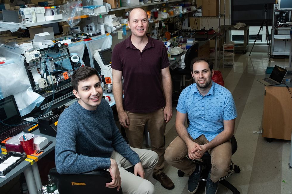
Rice University bioengineers (from left) Bagrat Grigoryan, Jordan Miller and Daniel Sazer and collaborators created a breakthrough bioprinting technique that could speed development of technology for 3D printing replacement organs and tissues. Photo by Jeff Fitlow/Rice University

Ad Placement 300x100
Ad Placement 300x600

CultureMap Emails are Awesome

Rice University lands $18M to revolutionize lymphatic disease detection

fresh funding

An arm of the U.S. Department of Health and Human Services has awarded $18 million to scientists at Rice University for research that has the potential to revolutionize how lymphatic diseases are detected and help increase survivability.

The lymphatic system is the network of vessels all over the body that help eliminate waste, absorb fat and maintain fluid balance. Diseases in this system are often difficult to detect early due to the small size of the vessels and the invasiveness of biopsy testing. Though survival rates of lymph disease have skyrocketed in the United States over the last five years, it still claims around 200,000 people in the country annually.

Early detection of complex lymphatic anomalies (CLAs) and lymphedema is essential in increasing successful treatment rates. That’s where Rice University’s SynthX Center, directed by Han Xiao and Lei Li, an assistant professor of electrical and computer engineering, comes in.

Aided by researchers from Texas Children’s Hospital, Baylor College of Medicine, the University of Texas at Dallas and the University of Texas Southwestern Medical Center, the center is pioneering two technologies: the Visual Imaging System for Tracing and Analyzing Lymphatics with Photoacoustics (VISTA-LYMPH) and Digital Plasmonic Nanobubble Detection for Protein (DIAMOND-P).

Simply put, VISTA-LYMPH uses photoacoustic tomography (PAT), a combination of light and sound, to more accurately map the tiny vessels of the lymphatic system. The process is more effective than diagnostic tools that use only light or sound, independent of one another. The research award is through the Advanced Research Projects Agency for Health (ARPA-H) Lymphatic Imaging, Genomics and pHenotyping Technologies (LIGHT) program, part of the U.S. HHS, which saw the potential of VISTA-LYMPH in animal tests that produced finely detailed diagnostic maps.

“Thanks to ARPA-H’s award, we will build the most advanced PAT system to image the body’s lymphatic network with unprecedented resolution and speed, enabling earlier and more accurate diagnosis,” Li said in a news release.

Meanwhile, DIAMOND-P could replace the older, less exact immunoassay. It uses laser-heated vapors of plasmonic nanoparticles to detect viruses without having to separate or amplify, and at room temperature, greatly simplifying the process. This is an important part of greater diagnosis because even with VISTA-LYMPH’s greater imaging accuracy, many lymphatic diseases still do not appear. Detecting biological markers is still necessary.

According to Rice, the efforts will help address lymphatic disorders, including Gorham-Stout disease, kaposiform lymphangiomatosis and generalized lymphatic anomaly. They also could help manage conditions associated with lymphatic dysfunction, including cancer metastasis, cardiovascular disease and neurodegeneration.

“By validating VISTA-LYMPH and DIAMOND-P in both preclinical and clinical settings, the team aims to establish a comprehensive diagnostic pipeline for lymphatic diseases and potentially beyond,” Xiao added in the release.

The ARPA-H award funds the project for up to five years.

Houston doctor wins NIH grant to test virtual reality for ICU delirium

Virtual healing

Think of it like a reverse version of The Matrix. A person wakes up in a hospital bed and gets plugged into a virtual reality game world in order to heal.

While it may sound far-fetched, Dr. Hina Faisal, a Houston Methodist critical care specialist in the Department of Surgery, was recently awarded a $242,000 grant from the National Institute of Health to test the effects of VR games on patients coming out of major surgery in the intensive care unit (ICU).

The five-year study will focus on older patients using mental stimulation techniques to reduce incidences of delirium. The award comes courtesy of the National Institute on Aging K76 Paul B. Beeson Emerging Leaders Career Development Award in Aging.

“As the population of older adults continues to grow, the need for effective, scalable interventions to prevent postoperative complications like delirium is more important than ever,” Faisal said in a news release.

ICU delirium is a serious condition that can lead to major complications and even death. Roughly 87 percent of patients who undergo major surgery involving intubation will experience some form of delirium coming out of anesthesia. Causes can range from infection to drug reactions. While many cases are mild, prolonged ICU delirium may prevent a patient from following medical advice or even cause them to hurt themselves.

Using VR games to treat delirium is a rapidly emerging and exciting branch of medicine. Studies show that VR games can help promote mental activity, memory and cognitive function. However, the full benefits are currently unknown as studies have been hampered by small patient populations.

Faisal believes that half of all ICU delirium cases are preventable through VR treatment. Currently, a general lack of knowledge and resources has been holding back the advancement of the treatment.

Hopefully, the work of Faisal in one of the busiest medical cities in the world can alleviate that problem as she spends the next half-decade plugging patients into games to aid in their healing.