The new collaborative hub will foster research into cell therapies, artificial intelligence, nanotechnologies, and more. Photo via tmc.edu

Two Houston organizations recently announced a new hub that will focus on developing cell therapies, nanotechnologies, cancer vaccines, artificial intelligence, and molecular imaging.

Rice University and The University of Texas MD Anderson Cancer Center have teamed up to “drive industry growth and advance life-saving technologies” through the newly established Cancer Bioengineering Collaborative, according to a news release announcing the initiative.

The collaboration between the two institutions includes fundamental and translational cancer research, developing new technologies for cancer detection and therapy, and securing external funding in support of further research and training.

Leading the hub will be Rice researcher and Cancer Prevention and Research Institute of Texas (CPRIT) scholar Gang Bao and MD Anderson’s Dr. Jeffrey Molldrem.

“There is tremendous potential in bringing together experts in engineering and cancer as part of this focused, collaborative framework that is truly unique, not only owing to the complementary nature of the respective strengths but also because this is the first formal joint research initiative of its kind between the two institutions,” says Bao, department chair and Foyt Family Professor of Bioengineering, professor of chemistry, materials science and nanoengineering and mechanical engineering, in the release.

The joint effort will also host monthly seminars focused on cancer bioengineering, annual retreats to highlight research and international leaders in cancer and bioengineering, and also a seed grant program to fund research projects in the early stages of development.

“From fundamental discoveries in cancer science, tumor immunology and patient care to innovative engineering advances in drug delivery systems, nanostructures and synthetic biology, there is great potential for enabling cross-disciplinary collaboration to develop new technologies and approaches for detecting, monitoring and treating cancer,” Molldrem, chair of Hematopoietic Biology & Malignancy at MD Anderson, says in the release. “Our goal is to bridge the gap between bioengineering and cancer research to create transformative solutions that significantly improve patient outcomes.”

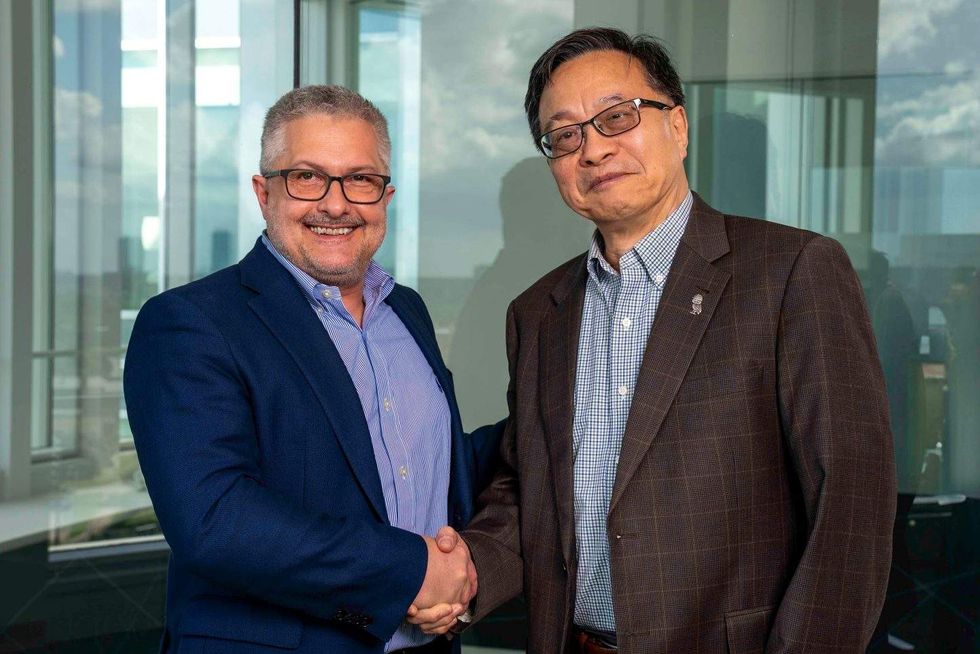
Dr. Jeff Molldrem (left) and Gang Bao will lead the new collaborative hub. Photo via MD Anderson

Ad Placement 300x100
Ad Placement 300x600

CultureMap Emails are Awesome

Rice University lands $18M to revolutionize lymphatic disease detection

fresh funding

An arm of the U.S. Department of Health and Human Services has awarded $18 million to scientists at Rice University for research that has the potential to revolutionize how lymphatic diseases are detected and help increase survivability.

The lymphatic system is the network of vessels all over the body that help eliminate waste, absorb fat and maintain fluid balance. Diseases in this system are often difficult to detect early due to the small size of the vessels and the invasiveness of biopsy testing. Though survival rates of lymph disease have skyrocketed in the United States over the last five years, it still claims around 200,000 people in the country annually.

Early detection of complex lymphatic anomalies (CLAs) and lymphedema is essential in increasing successful treatment rates. That’s where Rice University’s SynthX Center, directed by Han Xiao and Lei Li, an assistant professor of electrical and computer engineering, comes in.

Aided by researchers from Texas Children’s Hospital, Baylor College of Medicine, the University of Texas at Dallas and the University of Texas Southwestern Medical Center, the center is pioneering two technologies: the Visual Imaging System for Tracing and Analyzing Lymphatics with Photoacoustics (VISTA-LYMPH) and Digital Plasmonic Nanobubble Detection for Protein (DIAMOND-P).

Simply put, VISTA-LYMPH uses photoacoustic tomography (PAT), a combination of light and sound, to more accurately map the tiny vessels of the lymphatic system. The process is more effective than diagnostic tools that use only light or sound, independent of one another. The research award is through the Advanced Research Projects Agency for Health (ARPA-H) Lymphatic Imaging, Genomics and pHenotyping Technologies (LIGHT) program, part of the U.S. HHS, which saw the potential of VISTA-LYMPH in animal tests that produced finely detailed diagnostic maps.

“Thanks to ARPA-H’s award, we will build the most advanced PAT system to image the body’s lymphatic network with unprecedented resolution and speed, enabling earlier and more accurate diagnosis,” Li said in a news release.

Meanwhile, DIAMOND-P could replace the older, less exact immunoassay. It uses laser-heated vapors of plasmonic nanoparticles to detect viruses without having to separate or amplify, and at room temperature, greatly simplifying the process. This is an important part of greater diagnosis because even with VISTA-LYMPH’s greater imaging accuracy, many lymphatic diseases still do not appear. Detecting biological markers is still necessary.

According to Rice, the efforts will help address lymphatic disorders, including Gorham-Stout disease, kaposiform lymphangiomatosis and generalized lymphatic anomaly. They also could help manage conditions associated with lymphatic dysfunction, including cancer metastasis, cardiovascular disease and neurodegeneration.

“By validating VISTA-LYMPH and DIAMOND-P in both preclinical and clinical settings, the team aims to establish a comprehensive diagnostic pipeline for lymphatic diseases and potentially beyond,” Xiao added in the release.

The ARPA-H award funds the project for up to five years.

Houston doctor wins NIH grant to test virtual reality for ICU delirium

Virtual healing

Think of it like a reverse version of The Matrix. A person wakes up in a hospital bed and gets plugged into a virtual reality game world in order to heal.

While it may sound far-fetched, Dr. Hina Faisal, a Houston Methodist critical care specialist in the Department of Surgery, was recently awarded a $242,000 grant from the National Institute of Health to test the effects of VR games on patients coming out of major surgery in the intensive care unit (ICU).

The five-year study will focus on older patients using mental stimulation techniques to reduce incidences of delirium. The award comes courtesy of the National Institute on Aging K76 Paul B. Beeson Emerging Leaders Career Development Award in Aging.

“As the population of older adults continues to grow, the need for effective, scalable interventions to prevent postoperative complications like delirium is more important than ever,” Faisal said in a news release.

ICU delirium is a serious condition that can lead to major complications and even death. Roughly 87 percent of patients who undergo major surgery involving intubation will experience some form of delirium coming out of anesthesia. Causes can range from infection to drug reactions. While many cases are mild, prolonged ICU delirium may prevent a patient from following medical advice or even cause them to hurt themselves.

Using VR games to treat delirium is a rapidly emerging and exciting branch of medicine. Studies show that VR games can help promote mental activity, memory and cognitive function. However, the full benefits are currently unknown as studies have been hampered by small patient populations.

Faisal believes that half of all ICU delirium cases are preventable through VR treatment. Currently, a general lack of knowledge and resources has been holding back the advancement of the treatment.

Hopefully, the work of Faisal in one of the busiest medical cities in the world can alleviate that problem as she spends the next half-decade plugging patients into games to aid in their healing.