Houston-based Dr. Theodoros Voloyiannis was one of six involved in a remote surgery in space demonstration. Photo courtesy of Texas Oncology

A small surgical robot at the International Space Station completed its first surgery demo in zero gravity last week, and one of the surgeons tasked with the remote robotic operations on simulated tissue was Houston-based Dr. Theodoros Voloyiannis.

Voloyiannis took part in what is being referred to as “surgery in space” by being one of the six doctors remotely controlling spaceMIRA — Miniaturized In Vivo Robotic Assistant — that performed several operations on simulated tissue at the lab located in the space station. The surgeons operated remotely from earth in Lincoln, Nebraska. The remote surgeons worked to control the robot's hands to provide tension to the simulated tissue made of rubber bands. They then used the other hand to dissect the elastic tissue with scissors.

“I said during the procedure ‘it was a small rubber band cut, but a great leap for surgery,’“ Voloyiannis tells InnovationMap. “This was a huge milestone for me personally in my career.”

The robot was developed by Virtual Incision Corporation, and made possible through a partnership between NASA and the University of Nebraska. The team of surgeons took part in a demonstration that is considered a common surgical task, as they dissected the correct piece of tissue under pressure.

Latency is the time delay between when the command is sent and the robot receives it, and that was the big challenge the team faced. The delay was about 0.85 of a second according to what the colorectal surgeon who worked on spaceMIRA Dr. Michael Jobst said to CNN. The demo overall was a success according to the team, and posed a new-found adrenaline rush due to the groundbreaking innovation.

“The excitement of the new and the unknown,” Voloyiannis says on the feeling of doing the first operation of its kind. “I never thought I’d be doing something like this when I was in training and in medical school.”

Voloyiannis serves as the chairman of colon and rectal surgery for The US Oncology Network. He was chosen for this experiment due to his experience and expertise performing robotic colorectal surgery. Voloyiannis and the developers are hopeful that this type of technology will soon allow doctors to perform this specialized robotic surgery on patients living in rural areas without a specialized surgeon nearby, military battlefields, as well as regularly in space one day.

“The same concept of remote surgery regularly in space could certainly be entertained,” Voloyiannis says. “When you do things with an absence of gravity and perform a surgery in that environment — of course that changes the way we do things. When you have an absence of gravity with bodily fluids, it is a very hard surgery, but with partial gravity that idea can be entertained.

"Remotely, internet connectivity would have to be considered and you’d have someone remote like me here, while potentially there you’d have someone with less training doing the procedure there guiding the robot," he continues. "It’s quite the concept though.”

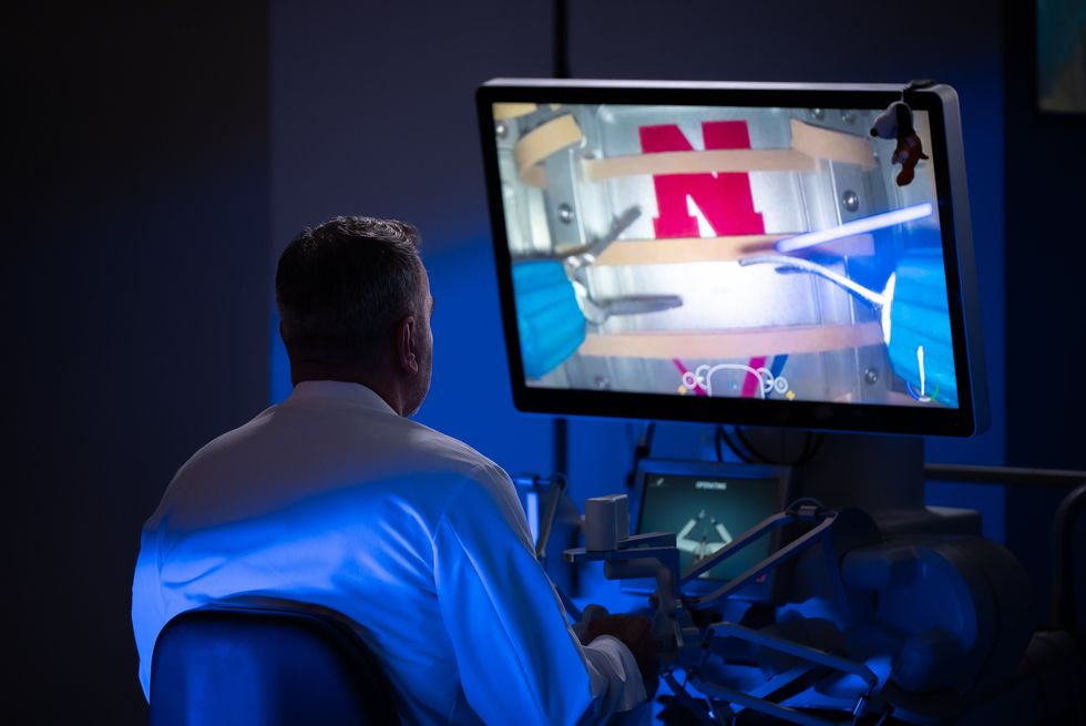
The doctors had to account for nearly a second of delay in connectivity. Photo courtesy of Texas Oncology

A mixed reality lab at the University of Houston is merging the physical and digital worlds. Photo via UH.edu

UH lab using mixed reality to optimize designs for the Moon and Mars

hi, tech

University of Houston researchers and students are bringing multiple realities together to help improve the design process for crewed space missions.

Helmed by Vittorio Netti, a researcher for UH and a space architect, the university has launched an XR Lab within the University of Houston architecture building. The lab allows researchers to combine mixed reality (MR), virtual reality (VR), augmented reality (AR) and extended reality (XR) to "blend the physical and digital worlds" to give designers a better understanding of life in space, according to a release from UH.

In the lab researchers can wear MY space suits and goggles, take a VR space walk, or feel what it's like to float to the International Space Station with the help of XR and a crane.

The area in which the researchers conduct this work is known as the "cage" and was developed during a six-month research and design study of lunar surface architecture sponsored by Boeing, which aimed to learn more about the design of a lunar terrain vehicle and a small lunar habitat.

The work is part of UH's Sasakawa International Center of Space Architecture (SICSA), which is led by Olga Bannova, a research associate professor and director of the space architecture graduate program at UH.

She says work like this will drastically cut down research and development time when designing space structures.

“These technologies should be harnessed to mitigate the dependency on physical prototyping of assets and help optimize the design process, drastically reducing research-and-development time and providing a higher level of immersion,” Bannova said in a statement.

Today the research team is shifting its focus on designing for a Mars landing. In the future, they aim to demonstrate and test the system for habitats designed for both lunar and Martian surfaces. They are also working with Boeing to test designs in microgravity, or zero gravity, which exists inside the International Space Station.

Mixed Reality Raising the Bar for Space Architecture on the Moon and MarsStep into this 'Cage' at the University of Houston where physical and digital worlds are merged, allowing students to see and ...

Ad Placement 300x100
Ad Placement 300x600

CultureMap Emails are Awesome

Rice University lands $18M to revolutionize lymphatic disease detection

fresh funding

An arm of the U.S. Department of Health and Human Services has awarded $18 million to scientists at Rice University for research that has the potential to revolutionize how lymphatic diseases are detected and help increase survivability.

The lymphatic system is the network of vessels all over the body that help eliminate waste, absorb fat and maintain fluid balance. Diseases in this system are often difficult to detect early due to the small size of the vessels and the invasiveness of biopsy testing. Though survival rates of lymph disease have skyrocketed in the United States over the last five years, it still claims around 200,000 people in the country annually.

Early detection of complex lymphatic anomalies (CLAs) and lymphedema is essential in increasing successful treatment rates. That’s where Rice University’s SynthX Center, directed by Han Xiao and Lei Li, an assistant professor of electrical and computer engineering, comes in.

Aided by researchers from Texas Children’s Hospital, Baylor College of Medicine, the University of Texas at Dallas and the University of Texas Southwestern Medical Center, the center is pioneering two technologies: the Visual Imaging System for Tracing and Analyzing Lymphatics with Photoacoustics (VISTA-LYMPH) and Digital Plasmonic Nanobubble Detection for Protein (DIAMOND-P).

Simply put, VISTA-LYMPH uses photoacoustic tomography (PAT), a combination of light and sound, to more accurately map the tiny vessels of the lymphatic system. The process is more effective than diagnostic tools that use only light or sound, independent of one another. The research award is through the Advanced Research Projects Agency for Health (ARPA-H) Lymphatic Imaging, Genomics and pHenotyping Technologies (LIGHT) program, part of the U.S. HHS, which saw the potential of VISTA-LYMPH in animal tests that produced finely detailed diagnostic maps.

“Thanks to ARPA-H’s award, we will build the most advanced PAT system to image the body’s lymphatic network with unprecedented resolution and speed, enabling earlier and more accurate diagnosis,” Li said in a news release.

Meanwhile, DIAMOND-P could replace the older, less exact immunoassay. It uses laser-heated vapors of plasmonic nanoparticles to detect viruses without having to separate or amplify, and at room temperature, greatly simplifying the process. This is an important part of greater diagnosis because even with VISTA-LYMPH’s greater imaging accuracy, many lymphatic diseases still do not appear. Detecting biological markers is still necessary.

According to Rice, the efforts will help address lymphatic disorders, including Gorham-Stout disease, kaposiform lymphangiomatosis and generalized lymphatic anomaly. They also could help manage conditions associated with lymphatic dysfunction, including cancer metastasis, cardiovascular disease and neurodegeneration.

“By validating VISTA-LYMPH and DIAMOND-P in both preclinical and clinical settings, the team aims to establish a comprehensive diagnostic pipeline for lymphatic diseases and potentially beyond,” Xiao added in the release.

The ARPA-H award funds the project for up to five years.

Houston doctor wins NIH grant to test virtual reality for ICU delirium

Virtual healing

Think of it like a reverse version of The Matrix. A person wakes up in a hospital bed and gets plugged into a virtual reality game world in order to heal.

While it may sound far-fetched, Dr. Hina Faisal, a Houston Methodist critical care specialist in the Department of Surgery, was recently awarded a $242,000 grant from the National Institute of Health to test the effects of VR games on patients coming out of major surgery in the intensive care unit (ICU).

The five-year study will focus on older patients using mental stimulation techniques to reduce incidences of delirium. The award comes courtesy of the National Institute on Aging K76 Paul B. Beeson Emerging Leaders Career Development Award in Aging.

“As the population of older adults continues to grow, the need for effective, scalable interventions to prevent postoperative complications like delirium is more important than ever,” Faisal said in a news release.

ICU delirium is a serious condition that can lead to major complications and even death. Roughly 87 percent of patients who undergo major surgery involving intubation will experience some form of delirium coming out of anesthesia. Causes can range from infection to drug reactions. While many cases are mild, prolonged ICU delirium may prevent a patient from following medical advice or even cause them to hurt themselves.

Using VR games to treat delirium is a rapidly emerging and exciting branch of medicine. Studies show that VR games can help promote mental activity, memory and cognitive function. However, the full benefits are currently unknown as studies have been hampered by small patient populations.

Faisal believes that half of all ICU delirium cases are preventable through VR treatment. Currently, a general lack of knowledge and resources has been holding back the advancement of the treatment.

Hopefully, the work of Faisal in one of the busiest medical cities in the world can alleviate that problem as she spends the next half-decade plugging patients into games to aid in their healing.